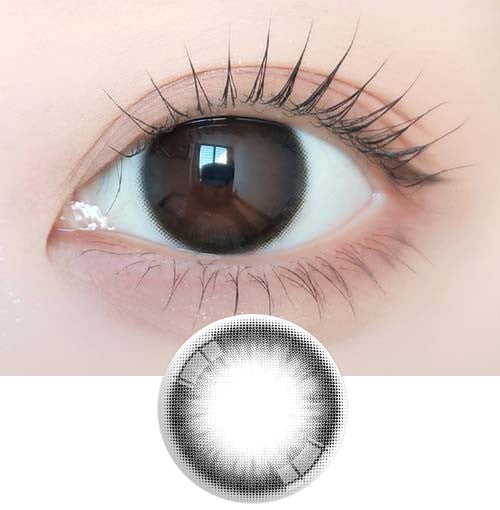
Eye Anatomy Explained: Key Parts and Functions
•Posted on April 28 2025
The human eye is an incredibly sophisticated and delicate organ that enables us to experience the world visually. Its structure is made up of multiple interconnected components, all working together seamlessly. The health of your eyes and the quality of your vision rely on each of these parts functioning properly.
In this guide, we'll explore the major parts of the eye and explain their key roles. We’ll also briefly describe how these structures collaborate to allow you to see.
Understanding the Eye’s Structure
(Imagine: Diagram showing a front view and a side view of the eye)
The eyeball itself is about the size of a ping-pong ball and includes both the external features you can see and the intricate internal structures you cannot. Internally, the eye is divided into three primary layers:
- Outer layer (sclera and cornea)
- Middle layer (choroid and ciliary body)
- Inner layer (retina)
To simplify things, we'll walk through the eye parts starting from the outermost surface and moving inward, roughly following the path light takes as it enters your eye.
Structures That Surround and Protect the Eye
The tissues and glands around the eyeball serve as protection and support for the sensitive internal parts. Important external structures include:
- Eyelids and Eyelashes: These work together to keep the eye surface moist, clear of debris, and shielded from bright light by reflexive blinking.
- Extraocular Muscles: These muscles anchor the eye within the orbital socket and control its movement in multiple directions—up, down, side to side, and diagonally.
- Lacrimal and Meibomian Glands: Located within the eyelids, these glands produce tears that maintain eye moisture, flush away irritants, and protect against infection.
The Eye’s Outer Layer: The Protective Shell
This fibrous outer coat includes parts that guard and maintain the eye’s shape:
- Sclera: The tough, white outer layer of the eyeball, rich in collagen fibers, providing structure and defense against injury.
- Cornea: A clear, dome-shaped covering over the front of the eye. It acts as a shield against dust, germs, and UV radiation, and plays an important role in focusing light.
- Conjunctiva: A thin, transparent membrane that lines the inside of the eyelids and covers the white part of the eyeball. When inflamed or infected, it results in a condition commonly known as conjunctivitis, or "pink eye."
The Middle Layer of the Eye: The Uvea
The middle layer of the eyeball, known as the uvea, contains several important structures that support blood flow and regulate the internal environment of the eye.
- Iris and Pupil: The iris is the colored part you see when looking into someone’s eyes. At its center lies the pupil, a dark circular opening that controls how much light enters the eye. The pupil changes size automatically—dilating (widening) in dim lighting and constricting (shrinking) in bright conditions to optimize vision.
- Ciliary Body: This small ring-shaped tissue produces aqueous humor, the clear fluid that nourishes the eye and maintains proper pressure. The fluid drains through a tiny passage known as the drainage angle; if fluid builds up too much, it can lead to elevated eye pressure, a key factor in glaucoma.
- Choroid: Lying between the retina and the sclera, the choroid is a thin vascular layer packed with blood vessels that deliver oxygen and nutrients to various parts of the eye.
The Inner Structures of the Eye: Vision's Control Center
The innermost layer at the back of the eye is made of specialized nerve tissue responsible for capturing light and transmitting visual information to the brain.
- Lens: Sitting just behind the iris, the lens is a transparent, flexible structure that focuses incoming light onto the retina, helping create a sharp image.
- Vitreous Body: The vitreous humor is a clear, gel-like material that fills the large space between the lens and the retina. It helps maintain the eye's rounded shape and also supports internal structures.
- Retina: Acting like the film in a camera, the retina captures light and converts it into neural signals. This complex tissue is essential for transforming visual stimuli into messages the brain can interpret.
- Macula: Found at the center of the retina, the macula is responsible for central vision, fine detail recognition, and color perception. Damage to this area, as seen in age-related macular degeneration (AMD), can severely impact daily activities like reading and recognizing faces.
- Optic Nerve: The optic nerve serves as the information highway between the retina and the brain, carrying the electrical impulses that allow us to perceive images.
How Vision Actually Happens
Although it feels effortless, vision is the result of an incredibly sophisticated process happening inside your eyes and brain. Each part of the eye plays a critical role in making sure you perceive depth, recognize objects, distinguish colors, and detect movement. Here’s a quick overview of how the human visual system works:
- Light enters the eye first through the cornea and passes through the pupil, which sits at the center of the iris.
- The cornea and lens both bend (or refract) the incoming light, directing it toward the retina at the back of the eye.
- The retina’s photoreceptors—specialized light-sensitive cells—convert the light into electrical signals.
- These signals travel along the optic nerve to the brain.
- The brain’s visual cortex processes and combines signals from both eyes, turning them into the clear, colorful images you experience every day.
(Imagine a simple diagram here showing the light pathway.)
Keeping Your Eyes—and Vision—Healthy
It’s clear that every structure inside the eye must work in harmony for you to see properly. Protecting your eyesight starts with maintaining the health of these parts, and the best way to do that is by scheduling a comprehensive eye exam every year.
If it's been a year or longer since your last exam—or if you’ve noticed any changes in your vision—now’s the time to book an appointment. With your new understanding of eye anatomy, you might even appreciate the check-up from a whole new perspective.


